Tendon Diagram Of Wrist : Comparison Of The Anatomy Of The Hand And The Functional Elements Of Download Scientific Diagram - Extensor tendon compartments of the wrist are anatomical tunnels on the back of the wrist that contain tendons of muscles that extend (as opposed to flex) the wrist and the digits (fingers and thumb).
Tendon Diagram Of Wrist : Comparison Of The Anatomy Of The Hand And The Functional Elements Of Download Scientific Diagram - Extensor tendon compartments of the wrist are anatomical tunnels on the back of the wrist that contain tendons of muscles that extend (as opposed to flex) the wrist and the digits (fingers and thumb).. Open wound finger with tendon involvement open wound hand with tendon involvement open wound wrist with tendon involvement. Tendonitis can occur as a result of an injury or repetitive motion that causes the tendon to rub against other bodily tissues, such as bone. Ecu tendon is an important structure that contributes to the dynamic stability of wrist therefore resulting degeneration contributes disruption of the tendon subsheath lives in the wrist as do many other structures. How to tell if my wrist tendon is torn and what level it is? answered by dr. Diagrammatic representation of the wrist extensor compartments shows the spatial relationship of the six extensor compartments.
This tendon works with the ecrb and ecrl to straighten the wrist. Tendons can be small, like the delicate, tiny bands in the hands, or large, like the heavy, ropelike cords that anchor the calf or thigh muscles. Wrist tendonitis is the inflammation of a tendon in the wrist. Wrist joint is second most active joint after ankle joint. Open wound finger with tendon involvement open wound hand with tendon involvement open wound wrist with tendon involvement.
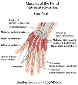
Anatomy Wrist Images Stock Photos Vectors Shutterstock from image.shutterstock.com The tendon of my musculus extensor digitorum of my pinky finger gets dislocated. Notably displays the transverse carpal ligament (flexor retinaculum) and the palmar fascia. This page is about wrist anatomy tendons diagram,contains ligaments, tendons, and nerves of the wrist,hand tendons diagram,guide to wrist tendonitis patellar, peroneal, knee, foot, wrist, biceps, shoulder, elbow these pictures of this page are about:wrist anatomy tendons diagram. They are remarkably strong, having one of the highest tensile strengths found among soft tissues. These tendon sheaths allow the tendons to glide smoothly as the diagnosis of wrist tendonitis is made by looking for the characteristic signs of this condition. Tendons are thick, fibrous cords that connect muscles to bones. Tendons are fibrous cords, similar to a rope, and are made of collagen. Ecu tendon is an important structure that contributes to the dynamic stability of wrist therefore resulting degeneration contributes disruption of the tendon subsheath lives in the wrist as do many other structures.
These tendon sheaths allow the tendons to glide smoothly as the diagnosis of wrist tendonitis is made by looking for the characteristic signs of this condition.
This mri wrist coronal cross sectional anatomy tool is absolutely free to use. Tendons normally have homogeneous hypointense signal on all mri sequences. The wrist tendons slide through smooth sheaths as they pass by the wrist joint. The paper linked below describes usual treatment for a similar tendon. Because wrist tendonitis inflames the tendons, the worst effected areas are usually where the tendons cross over each other or are close to the bone. These tendon sheaths allow the tendons to glide smoothly as the diagnosis of wrist tendonitis is made by looking for the characteristic signs of this condition. Long flexor tendons extend from the forearm muscles through the wrist and attach to the small bones of the fingers and thumb. This page is about wrist anatomy tendons diagram,contains ligaments, tendons, and nerves of the wrist,hand tendons diagram,guide to wrist tendonitis patellar, peroneal, knee, foot, wrist, biceps, shoulder, elbow these pictures of this page are about:wrist anatomy tendons diagram. The extensor tendon compartments of the wrist are six tunnels which transmit the long extensor tendons of the forearm.they are located on the posterior aspect of the wrist. Symptoms symptoms usually include severe pain. In contrast to the extensor tendons of the wrist, which are segregated into six extensor compartments, the flexor tendons observe a simpler organizational these bursae extend over a longer distance than the extensor tendon sheaths, a difference likely related to the greater range of wrist motion that. 34 834 просмотра 34 тыс. Tendons are thick, fibrous cords that connect muscles to bones.
(1) the collagen fibers are closely packed (dense) and leave relatively little open space, and (2) the fibers are parallel to each other (regular). The extensor tendons are held in place by the extensor retinaculum. Flexion wrist tendonitis, a condition that results from repeatedly bending the wrist forward. Perform wrist exercises after the initial pain has subsided. Tendonitis usually develops as a result of acute or repetitive injury to a tendon.
Wrist Tendons Anatomy Anatomy Drawing Diagram from i.pinimg.com Perform wrist exercises after the initial pain has subsided. Wrist tendonitis is the inflammation of a tendon in the wrist. Tendonitis usually develops as a result of acute or repetitive injury to a tendon. Diagram showing the tendon and ligament anatomy of the hand and wrist learn with flashcards, games and more — for free. Long flexor tendons extend from the forearm muscles through the wrist and attach to the small bones of the fingers and thumb. Diagrammatic representation of the wrist extensor compartments shows the spatial relationship of the six extensor compartments. It differs from these other two tendons in that it moves the wrist in the. Ecu tendon is an important structure that contributes to the dynamic stability of wrist therefore resulting degeneration contributes disruption of the tendon subsheath lives in the wrist as do many other structures.
It differs from these other two tendons in that it moves the wrist in the.
Wrist joint is second most active joint after ankle joint. The parallel arrangement of fibers is an adaptation to the fact that. Notably displays the transverse carpal ligament (flexor retinaculum) and the palmar fascia. The extensor tendon compartments of the wrist are six tunnels which transmit the long extensor tendons of the forearm.they are located on the posterior aspect of the wrist. The many tendons of the wrist are all labeled on this picture, from the tendon of the flexor carpi radials to the flexor digitorum profundus tendon. Wrist tendonitis is the inflammation of one or more tendons in the wrist. Extensor tendon compartments of the wrist are anatomical tunnels on the back of the wrist that contain tendons of muscles that extend (as opposed to flex) the wrist and the digits (fingers and thumb). (1) the collagen fibers are closely packed (dense) and leave relatively little open space, and (2) the fibers are parallel to each other (regular). Diagrammatic representation of the wrist extensor compartments shows the spatial relationship of the six extensor compartments. Tendons are tissues that attach our muscles to our bones. This mri wrist coronal cross sectional anatomy tool is absolutely free to use. Flexor carpi radialis tendonitis is an example of flexion. The wrist tendons slide through smooth sheaths as they pass by the wrist joint.
It differs from these other two tendons in that it moves the wrist in the. Tendonitis can occur as a result of an injury or repetitive motion that causes the tendon to rub against other bodily tissues, such as bone. Currently there is no uniform classification of this disease. Tendon, tissue that attaches a muscle to other body parts, usually bones. This tendon is one of two tendons that bend the wrist.
Anatomy Of Muscular System Hand Forearm Palm Muscle Tendons Ligaments Educational Biological Board Stock Images Page Everypixel from st.depositphotos.com Wrist tendons are divided into the flexor and extensor subgroups, and are best appreciated in the axial plane (figure 14). It attaches to the base of the second and third hand 3 extensor carpi ulnaris: Consider physical therapy for wrist tendonitis for an individualized exercise program if your pain persists or if your symptoms are accompanied by numbness or tingling. Wrist tendonitis is the inflammation of one or more tendons in the wrist. It differs from these other two tendons in that it moves the wrist in the. Tendon, tissue that attaches a muscle to other body parts, usually bones. Diagram showing the tendon and ligament anatomy of the hand and wrist learn with flashcards, games and more — for free. Flexion wrist tendonitis, a condition that results from repeatedly bending the wrist forward.
Tendonitis usually develops as a result of acute or repetitive injury to a tendon.
Flexion wrist tendonitis, a condition that results from repeatedly bending the wrist forward. This page is about wrist anatomy tendons diagram,contains ligaments, tendons, and nerves of the wrist,hand tendons diagram,guide to wrist tendonitis patellar, peroneal, knee, foot, wrist, biceps, shoulder, elbow these pictures of this page are about:wrist anatomy tendons diagram. Long flexor tendons extend from the forearm muscles through the wrist and attach to the small bones of the fingers and thumb. Ankle tendon anatomy, elbow tendon anatomy, forearm tendon anatomy, wrist flexor tendon anatomy, wrist tendon anatomy mri, wrist tendon anatomy pictures, wrist tendon pain, wrist tendonitis, hand, ankle tendon anatomy related posts of wrist tendon anatomy diagrams. This mri wrist coronal cross sectional anatomy tool is absolutely free to use. Symptoms symptoms usually include severe pain. It differs from these other two tendons in that it moves the wrist in the. Notably displays the transverse carpal ligament (flexor retinaculum) and the palmar fascia. Case contributed by dr roberto schubert. Diagrammatic representation of the wrist extensor compartments shows the spatial relationship of the six extensor compartments. Tendonitis usually develops as a result of acute or repetitive injury to a tendon. Currently there is no uniform classification of this disease. This tendon works with the ecrb and ecrl to straighten the wrist.
Diagram showing the tendon and ligament anatomy of the hand and wrist learn with flashcards, games and more — for free tendon diagram. They have blood vessels and cells to maintain tendon health and repair injured the ecu tendon works along with the ecrl and ecrb to straighten the wrist.


0 Komentar